Leishmaniosis in Cats
Most FeL case reports are from European and Mediterranean endemic areas where the number of pet cats is high. However, FeL remains rare, as a disease, even in areas where leishmaniosis is common in dogs and feline infection is frequent. It is postulated that cats are therefore less susceptible than dogs to L. infantum infection. However, it cannot be excluded that the disease is still underdiagnosed because it is neglected – or even ignored – by many practitioners, and clinical suspicion is challenging when suggestive signs are lacking, or the clinical presentation is overlooked by signs of a concurrent disease. Moreover, in some endemic areas, the level of cat medical care is generally lower compared to dogs.
About 140 clinical cases have been reported in Europe during the last 3 decades (Italy, France, Spain and Portugal) with some cases diagnosed in Germany, Switzerland and the UK in cats imported from endemic regions.
Host factors predisposing to susceptibility probably exist, as roughly half of the reported clinical cases have been observed in cats that could have had an impaired immune system secondary to FIV or feline leukemia virus (FeLV) or dual FIV and FeLV coinfections, long-term immune-suppressive therapies, malignant neoplasia, or debilitating diseases. However, the level of evidence of this observation is low as it is not supported by controlled studies.
The disease has been diagnosed in adult cats (2-21 years) with a median age of 9 years.
The incubation period can last years as suggested by clinical manifestations observed in non-endemic areas several years after the relocation of cats from endemic areas.
The course of disease is mostly chronic and progressive.
Similar to canine leishmaniosis, cutaneous and mucocutaneous lesions and lymphadenomegaly are the most frequently reported manifestations (Table 1). Ulcers and nodules are the most common cutaneous and mucocutaneous lesions observed, mainly distributed on the head or on distal limbs (Figures 2-8). Hemorrhagic nodules are atypical skin lesions (Figure 9).
Uveitis is a frequent ocular lesion (Figure 10) in FeL. Oral lesions consist of nodules (tongue and gingival mucosa).
In recent years, atypical presentations have been reported including upper respiratory tract disease manifested with chronic nasal discharge and severe obstructive syndrome in case of granulomatous inflammation with nodular or infiltrative masses. Other single cases with atypical presentations where the causative role of L. infantum has been documented with presentations of chronic diarrhea, mastitis and cutaneous panniculitis.
The clinicopathological evaluation of cats with leishmaniosis shows hyperglobulinemia with hypergammaglobulinemia frequently (Figure 10), mild normocytic normochromic non-regenerative anemia, and proteinuria.
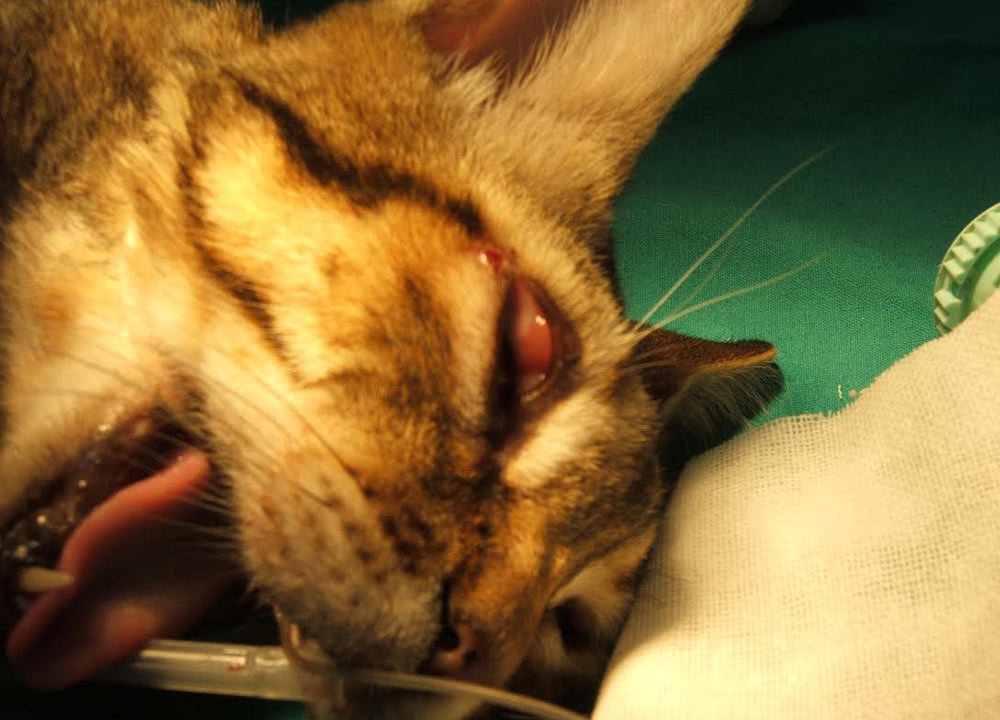
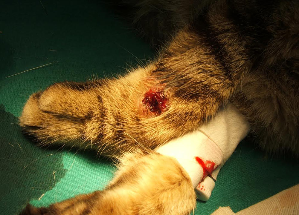
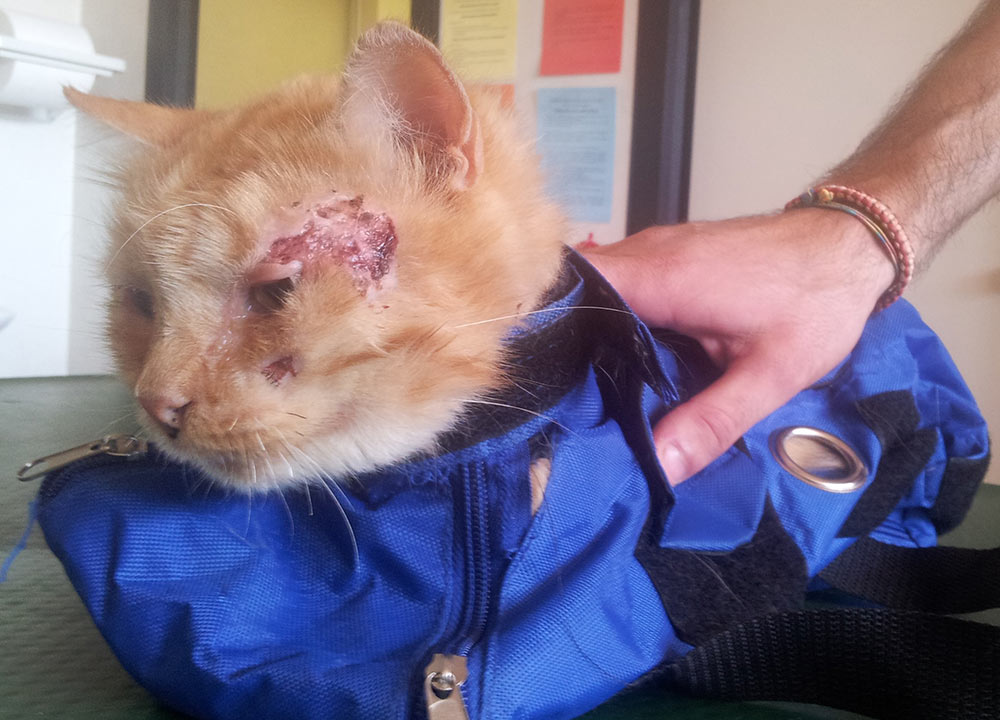
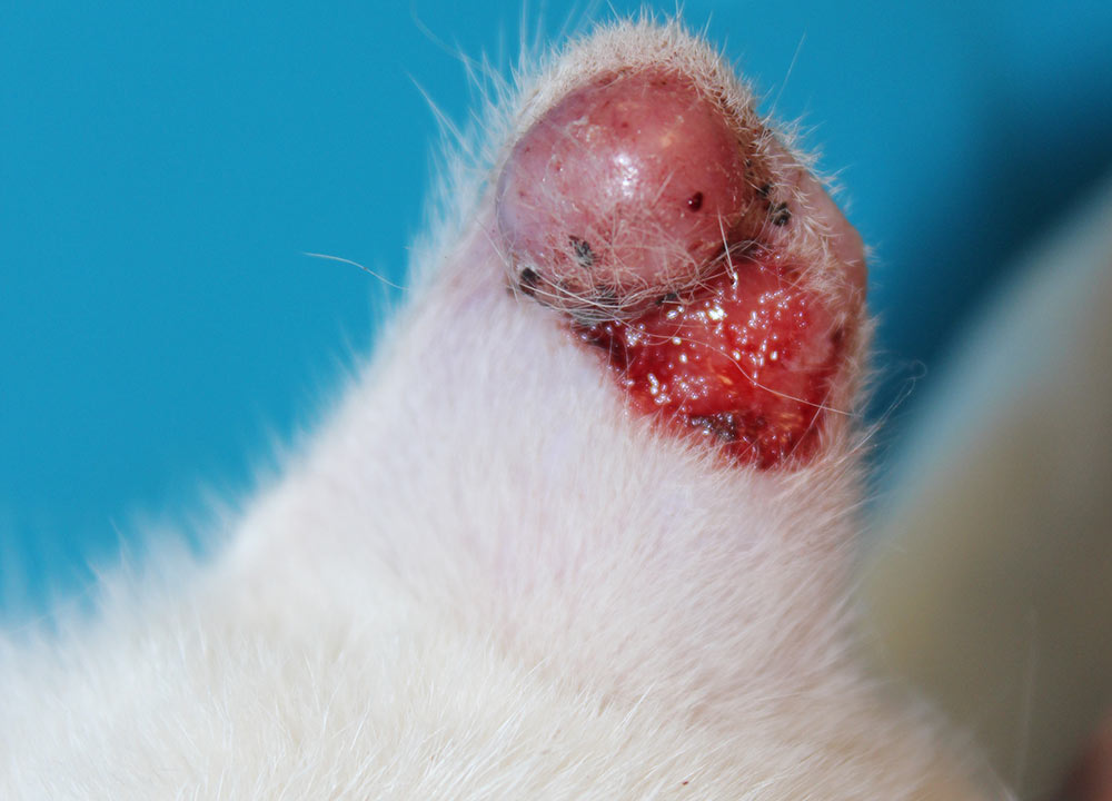
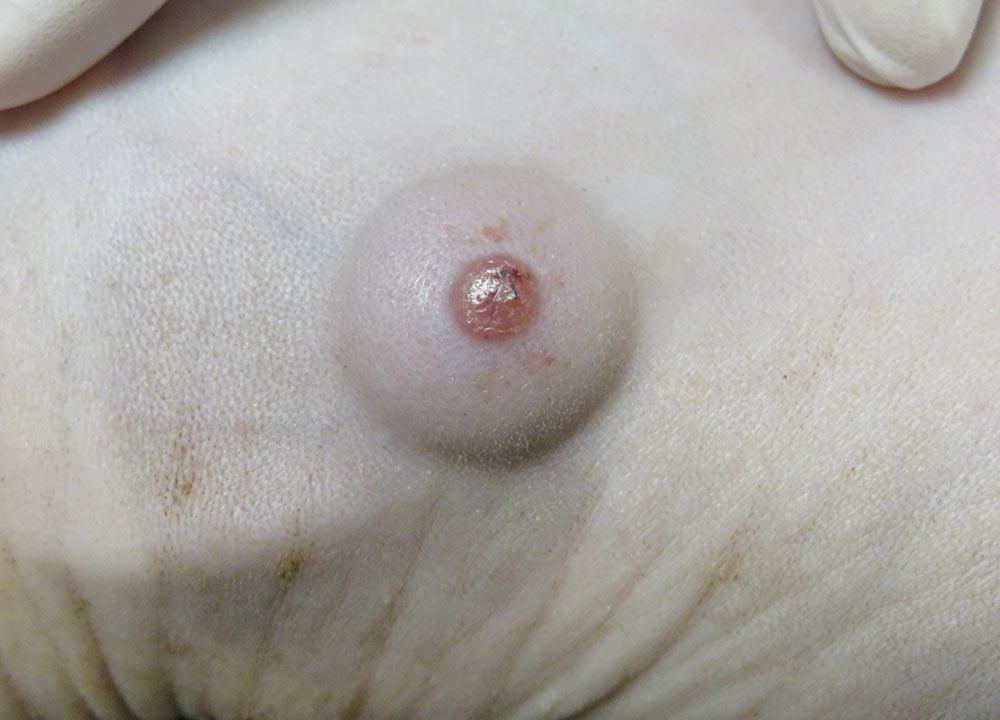
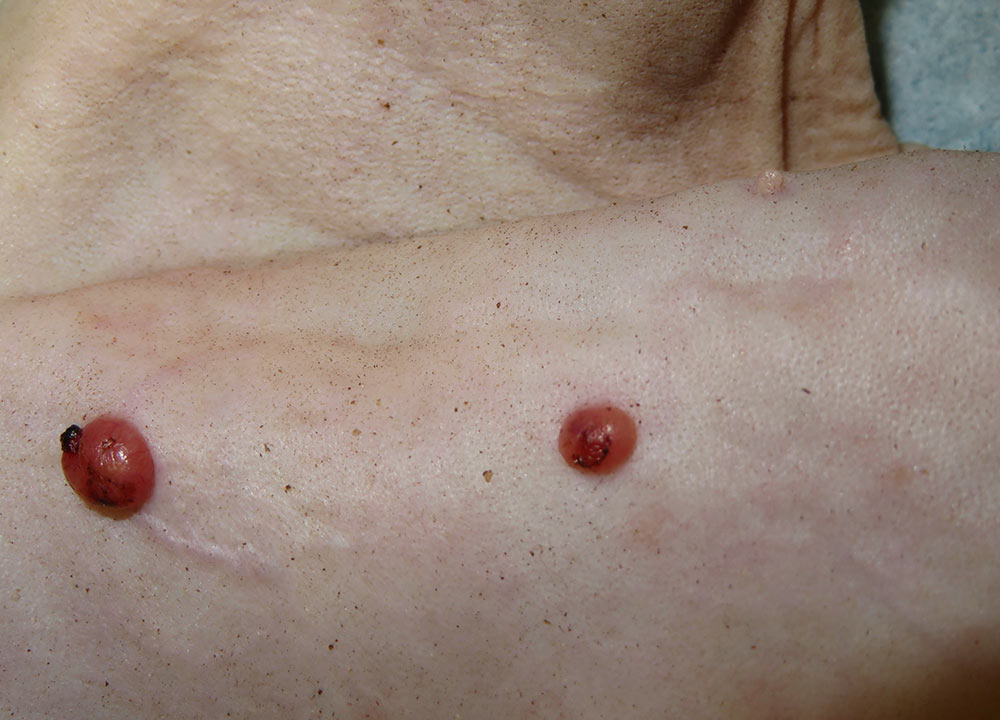
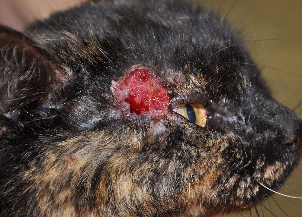
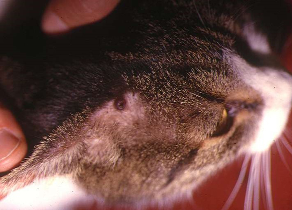
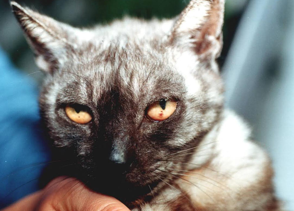
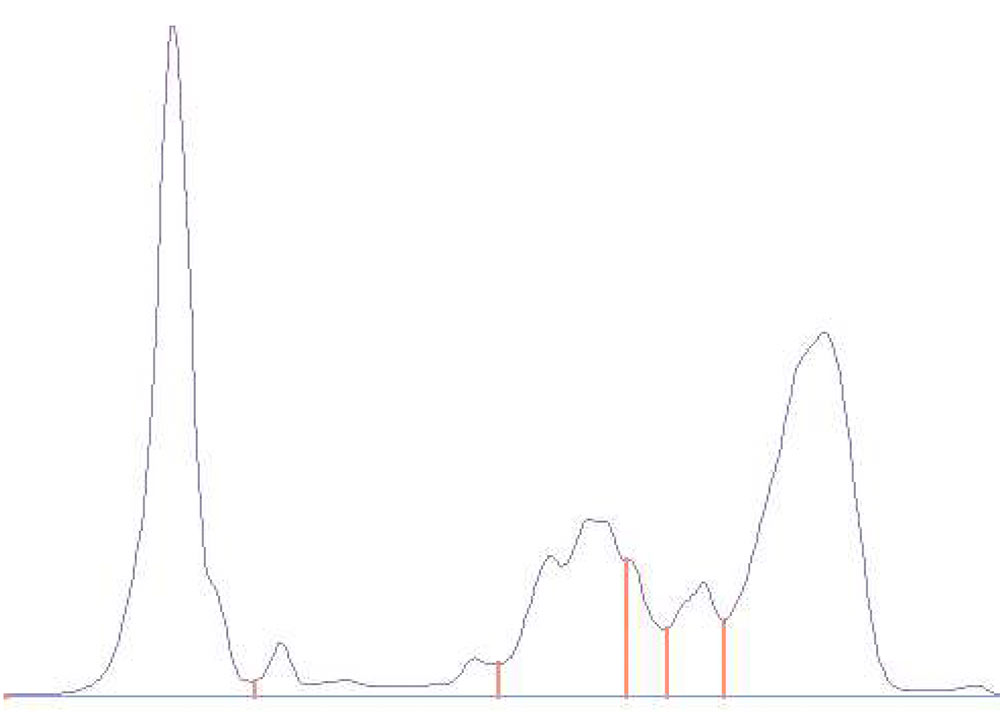
Table 1. Clinical and clinicopathological abnormalities reported in FeL (listed in decreasing order of frequency)
| CLINICAL FINDINGS | CLINICOPATHOLOGICAL ABNORMALITIES |
| Frequent* | |
|
Skin and/or mucocutaneous lesions Lymphadenomegaly |
Hyperglobulinemia Hypergammaglobulinemia |
| Uncommon** | |
|
Ocular lesions Oral lesions General signs (lethargy, anorexia,weight loss) |
Proteinuria Mild-to-moderate non-regenerative anemia |
| Rare*** | |
|
Pale/icteric mucous membranes Splenomegaly and/or hepatomegaly Chronic nasal discharge Upper airway obstructive syndrome Cachexia Polyuria/Polydipsia Fever Diarrhea Mastitis Subcutaneous panniculitis Abortion |
Azotemia Hypoalbuminemia Monocytosis Neutrophilia Pancytopenia |
* around 50% of cases
** around 30% of cases
*** less than 25% of cases