Diagnostic Challenges in the Era of Canine Leishmania infantum Vaccines
Diagnostic Challenges in the Era of Canine Leishmania infantum Vaccines
Solano-Gallego L, Cardoso L, Pennisi MG, Petersen Ch, Bourdeau P, Oliva G, Miró G, Ferrer Ll, Baneth G
(Trends in Pararasitology 2017, 33(9): 706-717)
The diagnosis of canine leishmaniosis (CanL) is complex due to its variable clinical manifestations and laboratory findings. The availability of vaccines to prevent CanL has increased the complexity of diagnosis, as serological tests may not distinguish between naturally infected and vaccinated dogs. Current practices of prevaccination screening are not sufficiently sensitive to detect subclinically infected dogs, resulting in the vaccination of infected animals, which may lead to disease in vaccinated dogs that are also infectious to sand flies. This review evaluates the current techniques for diagnosing CanL, and focuses on new challenges raised by the increasing use of vaccines against this disease. Important gaps in knowledge regarding the diagnosis of CanL are underscored to highlight the need for novel diagnostic test development.
6th World Congress on Leishmaniasis
Pilar Vidueira
LeishVet Continuing Education Programme
6th World Congress on Leishmaniasis
16-20 May 2017
Toledo, Spain
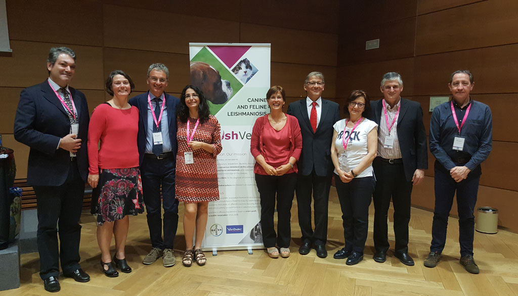
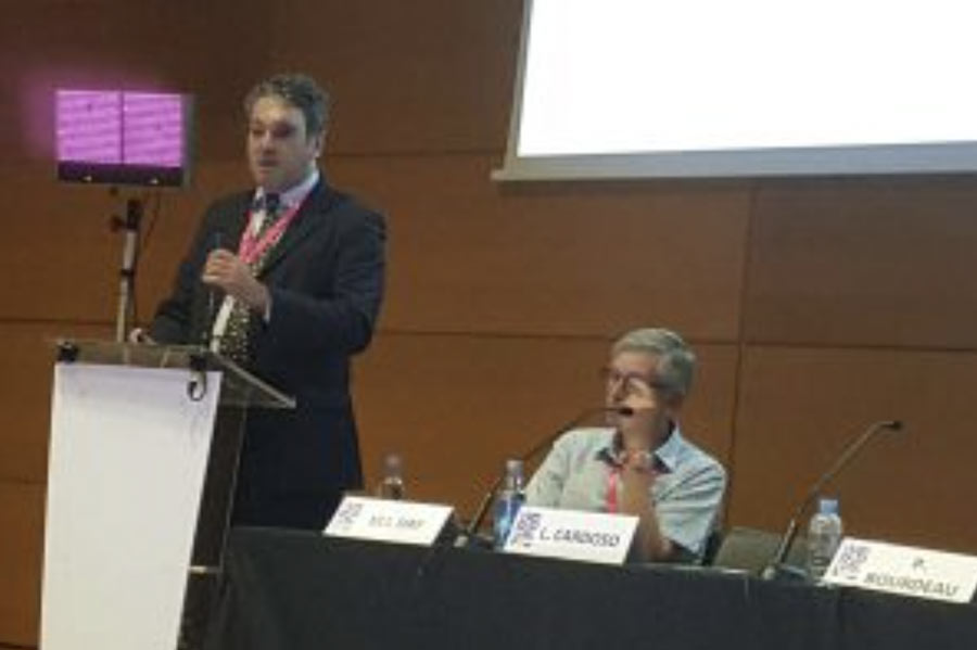
LeishVet Symposium – Toledo, May 2017
Coinciding with WORLDLEISH, the 6th World Congress on Leishmainasis celebrated in Toledo last May, the LeishVet group hold a special Symposium for clinical veterinaries on Saturday May 20th, sponsored by Bayer Health Care. Virbac also collaborated with the printing of the new version of the LeishVet Guidelines that was distributed to all attendees in English and Spanish.
The act was inaugurated by Dr. Jorge Alvar (Chair of the Congress) and Dr. Guadalupe Miró both acting as moderators during the event. The speakers were all the members of the LeishVet group with a special guest in Prof. Michael Day from Bristol University (UK) who gave a conference on leishmaniosis from One Health’s perspective. There was a total of 350 veterinaries attending the event all satisfied with the scientific programme (available for downloading above) and that actively participated in an interesting debate.


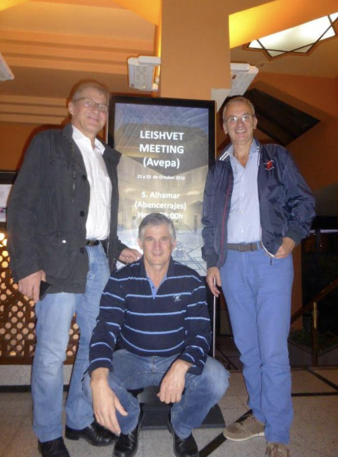
SEVC - X Southern European Veterinary Conference
Pilar Vidueira
LeishVet Continuing Education Programme
SEVC – X Southern European Veterinary Conference
20-22 October 2016
Granada, Spain
8th World Congress of Veterinary Dermatology
Pilar Vidueira
LeishVet Continuing Education Programme
8th World Congress of Veterinary Dermatology
4 June 2016
Bordeaux, France
Chairperson: Patrick Bourdeau
Attendance: Approximately 130 veterinarians
North America current situation
Speaker: Christine Petersen
New reservoirs & new species transmission
Speaker: Gad Baneth
What’s new in immunology of CanL
Speaker: Christine Petersen
What’s new in skin interface? Use of antiallergic and immunosuppressive drugs in Leishmania infected dogs
Speakers: Patrick Bourdeau and Lluís Ferrer
What’s new on clinical aspects? New Leishmania species in dogs. Feline, horse leish clinical cases
Speakers: Gad Baneth and Maria Grazia Pennisi
What’s new on diagnostic approach?
Speakers: Laia Solano-Gallego and Luis Cardoso
Updated LeishVet recommendations for prevention
Speakers: Guadalupe Miró and Gaetano Oliva
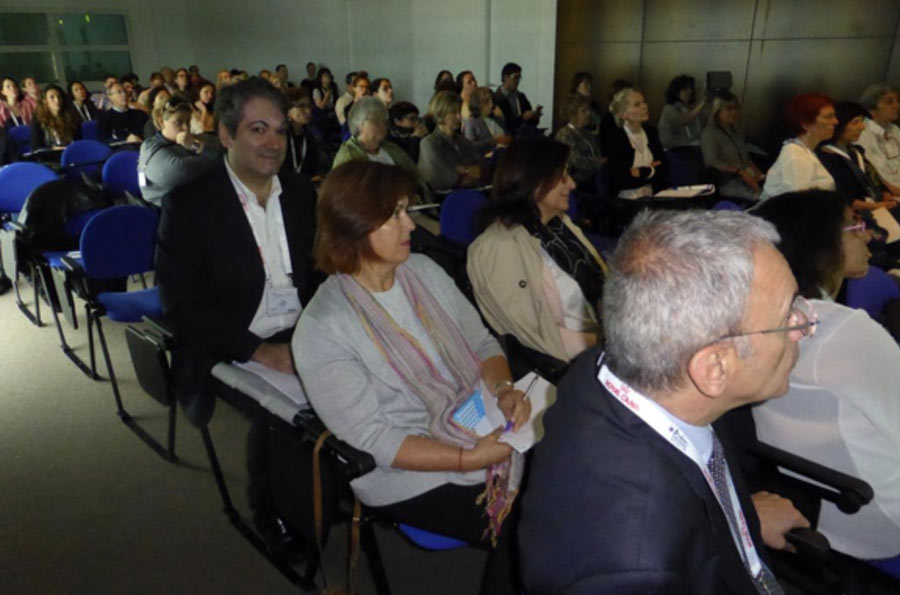

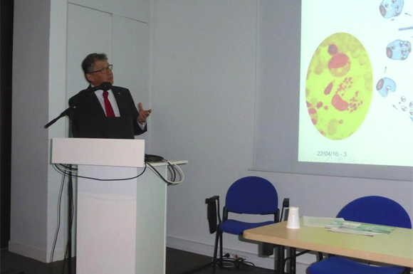
SCIVAC International Congress
Pilar Vidueira
LeishVet Continuing Education Programme
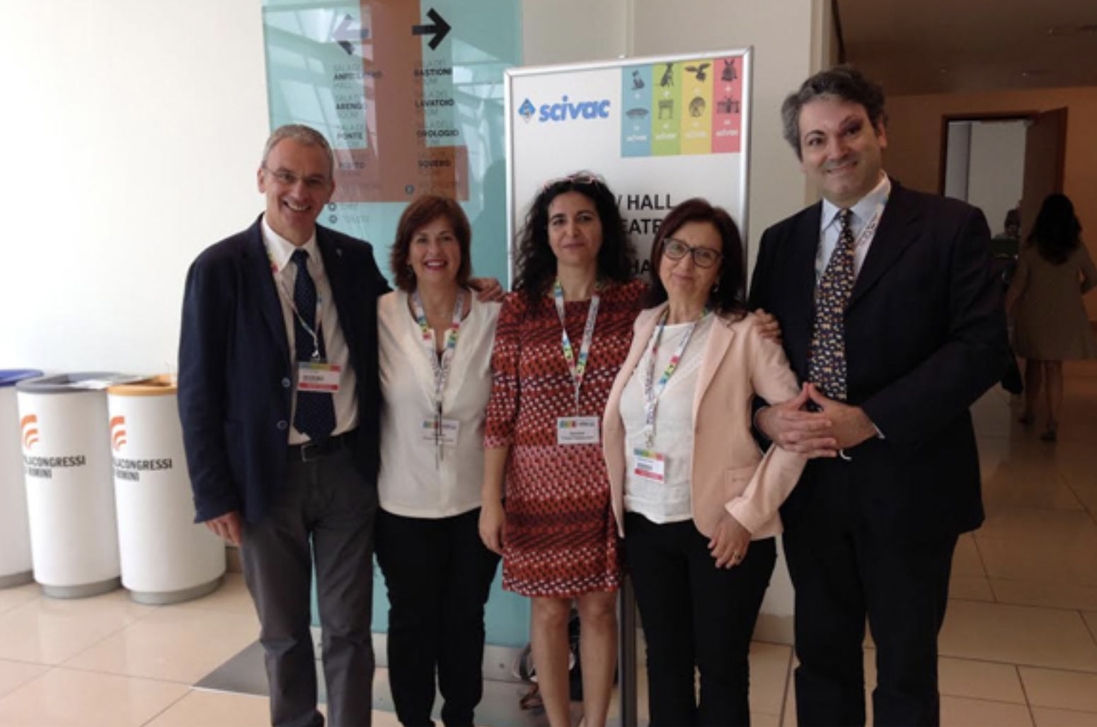
91º International SCIVAC Congress in collaboration with Zoomark
28 May 2016
Palacongressi della Riviera di Rimini, Italy
Tittle: “Update on epidemiology, Diagnosis and Control of Canine and Feline Leishmaniosis”
Chairperson: Gaetano Oliva
Attendance: Approximately 250 veterinarians
Dynamics in Europe. New reservoirs & new species
Speaker: Luis Cardoso
Diagnosis in the era of vaccines
Speaker: Laia Solano-Gallego
Feline leishmaniosis: update and clinical cases
Speaker: Maria Grazia Pennisi
Prevention
Speaker: Gaetano Oliva and Guadalupe Miró


6th World Congress on Leishmaniasis
Pilar Vidueira
The WHO ColIaborating Centre for Leishmaniasis at the Instituto de Salud Carlos III, Madrid and the Drugs for Neglected Diseases initiative, Geneva have joined efforts to co-organize the WorldLeish-6 Congress. On behalf of the WorldLeish Committee, it is agreat pleasure and privilege to invite you to the WorldLeish-6 Congress to be held in Toledo, Spain, from 16 to 20 on May 2017.
LeishVet update and recommendations on feline leishmaniosis
Pilar Vidueira
Limited data is available on feline leishmaniosis (FeL) caused by Leishmania infantum worldwide. The LeishVet group presents in this report a review of the current knowledge on FeL, the epidemiological role of the cat in L. infantum infection, clinical manifestations, and recommendations on diagnosis, treatment and monitoring, prognosis and prevention of infection, in order to standardize the management of this disease in cats. The consensus of opinions and recommendations was formulated by combining a comprehensive review of evidence-based studies and case reports, clinical experience and critical consensus discussions. While subclinical feline infections are common in areas endemic for canine leishmaniosis, clinical illness due to L. infantum in cats is rare. The prevalence rates of feline infection with L. infantum in serological or molecular-based surveys range from 0% to more than 60%. Cats are able to infect sand flies and, therefore, they may act as a secondary reservoir, with dogs being the primary natural reservoir. The most common clinical signs and clinicopathological abnormalities compatible with FeL include lymph node enlargement and skin lesions such as ulcerative, exfoliative, crusting or nodular dermatitis (mainly on the head or distal limbs), ocular lesions (mainly uveitis),feline chronic gingivostomatitis syndrome, mucocutaneous ulcerative or nodular lesions, hypergammaglobulinaemia and mild normocytic normochromic anaemia. Clinical illness is frequently associated with impaired immunocompetence, as in case of retroviral coinfections or immunosuppressive therapy. Diagnosis is based on serology, polymerase chain reaction (PCR), cytology, histology, immunohistochemistry (IHC) or culture. If serological testing is negative or low positive in a cat with clinical signs compatible with FeL, the diagnosis of leishmaniosis should not be excluded and additional diagnostic methods (cytology, histology with IHC, PCR, culture) should be employed. The most common treatment used is allopurinol. Meglumine antimoniate has been administered in very few reported cases. Both drugs are administered alone and most cats recover clinically after therapy. Follow-up of treated cats with routine laboratory tests, serology and PCR is essential for prevention of clinical relapses. Specific preventative measures for this infection in cats are currently not available.

Fifth World Congress on Leishmaniasis (WorldLeish 5)
Pilar Vidueira
LeishVet Symposium
13-17 May 2013
Porto de Galinhas, Pernambuco, Brazil
Visit website
LeishVet guidelines for the practical management of canine leishmaniosis
Pilar Vidueira
Solano-Gallego L, Miró G, Koutinas A, Cardoso L, Pennisi MG, Ferrer L, Bourdeau P, Oliva G, Baneth G, The LeishVet Group
(Parasit Vectors 2011, 0;4:86)
The LeishVet group has formed recommendations designed primarily to help the veterinary clinician in the management of canine leishmaniosis. The complexity of this zoonotic infection and the wide range of its clinical manifestations, from inapparent infection to severe disease, make the management of canine leishmaniosis challenging. The recommendations were constructed by combining a comprehensive review of evidence-based studies, extensive clinical experience and critical consensus opinion discussions. The guidelines presented here in a short version with graphical topic displays suggest standardized and rational approaches to the diagnosis, treatment, follow-up, control and prevention of canine leishmaniosis. A staging system that divides the disease into four stages is aimed at assisting the clinician in determining the appropriate therapy, forecasting prognosis, and implementing follow-up steps required for the management of the leishmaniosis patient.
Directions for the diagnosis, clinical staging, treatment and prevention of canine leishmaniosis
Pilar Vidueira
Solano-Gallego L, Koutinas A, Miró G, Cardoso L, Pennisi MG, Ferrer L, Bourdeau P, Oliva G, Baneth G
(Vet Parasitol. 2009, 165(1-2):1-18)
Canine leishmaniosis (CanL) due to Leishmania infantum is a life threatening zoonotic disease with a wide distribution in the Old and New World and increasing importance also in non-endemic areas. The purpose of this report is to present a consensus of opinions on the diagnosis, treatment, prognosis and prevention of CanL in order to standardize the management of this infection. CanL is a disease in which infection far exceeds clinical disease due to the high prevalence of subclinical infection among endemic canine populations. The most useful diagnostic approaches include serology by quantitative techniques and PCR. High antibody levels are associated with severe parasitism and disease and are diagnostic of clinical leishmaniosis. However, the presence of lower antibody levels is not necessarily confirmatory for the disease and further work-up is necessary to confirm CanL by other diagnostic methods such as cytology, histopathology and PCR. We propose a system of four clinical stages, based on clinical signs, clinicopathological abnormalities and serological status. Suitable therapy and expected prognosis are presented for each of the stages. The combination of meglumine antimoniate and allopurinol is the first line therapeutic protocol. However, although most dogs recover clinically after therapy, complete elimination of the parasite is usually not achieved and infected dogs may eventually relapse. Follow-up of treated dogs with blood counts, serum biochemistry, urinalysis, serology and PCR is essential for prevention of relapses. Protection against sand fly bites by topical insecticides is effective in reducing infection, and recent development of vaccines has indicated that prevention by vaccination is feasible.
N.B.: for a reprint of the article please contact leishvet@ucm.es